Tag Archive for: genetic changes

How Is High-Risk Myeloma Assessed?

How Is High-Risk Myeloma Assessed? from Patient Empowerment Network on Vimeo.

Expert Dr. Jeffrey Matous explains how myeloma risk is determined, including staging, genetic analysis, and discusses the frequency of high-risk myeloma in patients. 

Dr. Jeffrey Matous is a myeloma specialist at the Colorado Blood Cancer Institute and the assistant chair in myeloma research for Sarah Cannon Research Institute. Learn more about Dr. Matous.

See More from Evolve Myeloma

Related Resources:

Questions and Considerations When Making Myeloma Treatment Decisions

Myeloma CAR T-Cell Therapy_ How Does It Work and What Are the Risks

Transcript:

Katherine:

You mentioned high-risk myeloma earlier. How do you determine if a patient is high risk or low risk?  

Dr. Jeffrey Matous:

Absolutely, so this is not uniformly agreed upon among myeloma doctors, but in general, we assess risk based on a few different things. One is called staging, and we stage myeloma unlike any other cancer, so it’s not staged like breast cancer, or lung cancer, or prostate cancer. It’s staged according to something called R-ISS, RISS, and you get, basically, a one, two, or a three.  

Those are your stages, and in general, if your stage III, you have higher risk disease, but even more than that, we’re beginning to understand how myeloma cells misbehave at the genetic level, and we know that there are certain genetic findings inside the myeloma cell that can convey higher risk features. It’s important to stress to patients that these are not genetic findings that they were born with or can pass on through hereditary. 

These are findings that occurred during the life of the patient that occurred by chance and developed inside that cell that turned into myeloma, and those are the genetic changes that we’re talking about. And we know that certain of these genetic changes confer higher risk disease. And in general, Katherine, if I see 100 people with myeloma, about 85 of the 100 will fall into what I call a standard risk category and about 15 percent will fall into what we call the high-risk category. 

Katherine:

Okay. That’s really good to know. Thank you.  

How is Multiple Myeloma Staged?

How is Multiple Myeloma Staged? from Patient Empowerment Network on Vimeo.

How is staging used in multiple myeloma? Watch as expert Dr. Abdullah Khan explains staging and its use, and myeloma patient and Empowerment Lead Lisa Hatfield shares how the use of staging and its factors have evolved over time.

Download Guide

Descargar Guía

See More from START HERE Myeloma

Related Resources:

What Are the Beginning Stages of Multiple Myeloma (MM)

What Are the Beginning Stages of Multiple Myeloma (MM)?

Where Should I START Following My Myeloma Diagnosis

Transcript:

Healthcare professionals use a combination of lab test results, imaging tests, and bone marrow exams to determine the stage of multiple myeloma. The revised International Staging System or Durie-Salmon Staging System may be used in determining a patient’s multiple myeloma stage. 

Dr. Abdullah Khan:

The patients are assigned stages I to III. To determine the ISS you need lab values for the beta-2 microglobulin and albumin. For the revised ISS, you add on the lab value for LDH, lactate dehydrogenase, and you also add in the chromosome risk profile. So, there are certain genetic changes that predict a more aggressive myeloma. And the ones added to the revised ISS staging system are translocation 4;14, translocation 14;16, and deletion 17p.” 

Lisa Hatfield:

So staging with multiple myeloma is really unique. A lot of times people think that every cancer has four stages. That is not true with myeloma. There are three stages for myeloma. And even now as of this year, some providers, some oncologists are not even staging myeloma. They’re looking at the risk factors, the cytogenetic risk factors to see if you’re standard risk or high risk. They’re not even using the staging system. It’s interesting. Back when I was diagnosed back in 2018, there were two staging systems used. One was the Durie-Salmon scale. I was diagnosed as stage III, which is the highest level. Mine was based on the fact that I had a lot of bone lesions and bone involvement.

But there’s also one called R-ISS, the revised ISS staging. And that one I was staged at stage I. So it’s really important to get a myeloma specialist on board quickly, because my myeloma specialist explained why I had two different stages I was staged at. She used the R-ISS, the more current system, the stage I and also looked at my cytogenetic risk factors. So your doctor will talk to you about FISH testing. And FISH testing might show something like translocations in the myeloma cells themselves or genetic abnormalities in the myeloma cells themselves. And that helps them dictate also what your risk will be going forward and how to treat that, how to treat your myeloma as a result of those risk factors and the stage you’re diagnosed at.

Your care provider may use other criteria in determining a person’s best optimal treatment. Make sure to ask if you have additional questions.

Understanding High-Risk DLBCL

Understanding High-Risk DLBCL from Patient Empowerment Network on Vimeo.

What is high-risk diffuse large B-cell lymphoma (DLBCL) exactly? Dr. Jane Winter explains progression of the disease, DLBCL subgroups, and treatment approaches that may bring high-risk DLBCL under control.

Dr. Jane Winter is a hematologist and medical oncologist at Robert H. Lurie Comprehensive Cancer Center at Northwestern University. More information on Dr. Winter here.

See More From The Pro-Active DLBCL Patient Toolkit

Related Programs:

Understanding Diffuse Large B-cell Lymphoma (DLBCL) and Its Subtypes

Understanding Diffuse Large B-cell Lymphoma (DLBCL) and Its Subtypes

What Are the Subtypes of DLBCL?

What Is Diffuse Large B-cell Lymphoma (DLBCL)?


Transcript:

Laura Beth:

Dr. Winter, what is high-risk DLBCL?   

Dr. Winter:

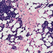
There are certain aggressive lymphomas that are more high-grade. And in recent years, we’ve identified a subset that has genetic changes in certain genes that lead to a very aggressive type of lymphoma that grows rapidly and is more difficult to cure with standard therapy. And what’s most important about identifying that subset is that it requires that at least most of our evidence is that the standard strategy that I just spoke about briefly in terms of what we use for transformed follicular lymphoma, the standard therapy we use for diffuse large B-cell lymphoma called R-CHOP where the “R” is standing for rituximab. 

Again, that antibody that targets a particular marker on the lymphoma cells, CD20 immunotherapy plus chemotherapy, CHOP chemotherapy which has been around for probably 50 years, this very standard backbone of treatment for diffuse large B-cell lymphoma is not sufficient. We don’t have head-to-head randomized phase three trials showing definitely that the more aggressive strategy is so much better, but most of the evidence is that standard R-CHOP just doesn’t do as well in curing these highly aggressive, and what we call high grade or double hit lymphomas.  

These are lymphomas that have these genetic changes, rearrangements in particular genes MYC and BCL2, and I like to think of it as MYC takes the brakes off of growth. So, it’s sort of the rapidly growing disease, and the BCL2 prevents the cells from dying. So, between that, you have a rapidly growing lymphoma that doesn’t stop. And so, that’s a special subgroup of diffuse large B-cell lymphoma. There are some other subcategories.  

Primary mediastinal, which tends to be a type of aggressive lymphoma that presents generally as a big mediastinal mass in the middle of the chest, usually younger patients, more often women than men, but still many, many men as well. So, high-risk generally for me means that it has a double hit. There’s MYC, M-Y-C, and BCL2 genetic changes. There are some other types, but that’s basically what I think of in terms of high-risk. When we think of garden-variety diffuse large B-cell lymphomas, there are some ways of categorizing patients into four different subcategories in terms of “risk.”   

The first of these was what we call the international prognostic index that was developed in the 70s based on patients who were enrolled on clinical trials. In those days, the diagnoses were probably not exactly what they are today in terms of how we categorize patients, but that is a set of clinical features that there’s a little app online and you can calculate whether a patient is high-risk, intermediate, low, low-intermediate risk. In this system remains fairly helpful, predictive. But, we have refined it significantly in modern times. 

And I have to say, not to toot my own horn, but one of my fellows, Dr. Zhao and I, a number of years ago using a database from the National Comprehensive Cancer Network, and patients who all treated with modern therapy and were diagnosed with modern techniques, we used that data to develop a new, improved international prognostic index. And this helps us better discriminate the four different categories, and it places greater weight on patient age, which is an important predictor, as well as the particular blood test result, the LDH, which is an enzyme that’s helpful in aggressive lymphomas and diffuse large B-cell lymphoma.  

And, this particular, what we call the NCCN-IPI, you can just put your age and LDH level and so on into this app and you just find it online, NCCN–IPI, and it will put patients into four different subgroups. And this predict outcomes and survival over a five-year period for these patients. It’s not hard and fast, like anything else, within every category. So, if you look at every patient in the low-risk group by the NCCN–IPI, there still are a few patients who will fail therapy, but the majority of patients will do very well.  

And, if you happen to be someone who is 80 years old with a high LDH and you fall into the high-risk patient group, the outcomes are not going to be as good, but that doesn’t mean every patient in that category fails treatment. So, there’s still gonna be some good outcomes. So, it’s helpful. These are guides, and they do help to identify patients who are, what we call, high-risk or high intermediate risk. So, this is another way of looking at predicting outcome apart from looking at whether a patient is what we call a “double hit” lymphoma. So, the double hit, I just want to make one point is that when a pathologist makes a diagnosis of diffuse large B-cell lymphoma, it is absolutely critical that that biopsy, the pathology be sent for a procedure called FISH.  

That stands for fluorescent in situ hybridization, and it’s a technique the pathologist can use, or special labs will use, to pull out, to identify those patients who have this very high-risk subset of lymphoma called the double hit with both a MYC and a BCL2 rearrangement. Whether a MYC and a BCL6 rearrangement falls into this same risk group, we’re deemphasizing that and really, it’s the double MYC and BLC2 group that’s the highest risk.  

How Do Myeloma Test Results Guide Prognosis and Treatment?

How Do Myeloma Test Results Guide Prognosis and Treatment? from Patient Empowerment Network on Vimeo.

Myeloma specialist Dr. Peter Forsberg explains how myeloma test results help in assessing the disease stage and prognosis, and how identification of chromosomal abnormalities may aid in treatment decisions.

Dr. Peter Forsberg is assistant professor of medicine at the University of Colorado School of Medicine and is a specialist in multiple myeloma. More about Dr. Forsberg here.

Download Program Resource Guide

See More From The Pro-Active Myeloma Patient Toolkit

Related Resources:

Why Myeloma Patients Should Speak Up: Advice from a Nurse Practitioner

Myeloma Treatment Decisions: What’s Right for You Resource Guide

Myeloma Targeted Therapy: Why Identifying Chromosomal Abnormalities Is Key

Transcript:

Katherine:                  

What do the results of these tests tell us about prognosis and treatment choices?

Dr. Forsberg:             

So, the tests that we do are important in terms of understanding some degree how aggressive the myeloma may be or what the prognosis may be. One of the most common or challenging things to break through when diagnosing myeloma or learning about your myeloma is that it’s a little different than other types of cancer. Unlike other cancers that’re more common, stage in myeloma is very different than it is in breast cancer or lung cancer or things that people may have more experience with. In myeloma, everybody has systemic disease.

That’s a part of the diagnosis of myeloma. It means it’s a body-wide condition. So, being stage I or II or III is very different than what it might be in other diseases where that has a huge prognostic impact and also, really shapes what treatment might be. In myeloma, we do use blood tests and chromosomal changes to help us assign a stage to the myeloma, which may tell us about how aggressive the myeloma may be over time.

But our treatment approaches tend to be pretty similar, even for people regardless of their stage. So, our goals are always to get patients’ myeloma under control and maintain it there. So, treatment ends up overlapping pretty substantially. Regardless of what those in initial tests are that stratify potential disease aggressiveness. That being said, there are some ways that we do adjust treatment potentially in patients that we see evidence of potentially more aggressive disease or less. And that might be ways that we amplify treatment regiments, adding extra medicines or using maintenance approaches that’re a little more robust to try to help overcome those high-risk features.

Katherine:                  

What about the significance of chromosomal abnormalities?

Dr. Forsberg:             

So, chromosomal abnormalities are part of some of those staging systems. They’re included in what we call our revised international staging system, as well as just being part of our routine risk assessment.

To try to understand myeloma. So, in myeloma, at this point those genetic changes or chromosomal changes don’t necessarily drive specific treatment choices except in that they may stratify how aggressive disease could be and may be informative in that regard.