Tag Archive for: skin cancer

Non-Melanoma Skin Cancer Treatment Options

Non-Melanoma Skin Cancer Treatment Options from Patient Empowerment Network on Vimeo.

What are the treatment options for advanced non-melanoma skin cancer? Dr. Sunandana Chandra shares insight on how a treatment approach is determined, the types of non-melanoma skin cancer, and discusses factors considered when choosing therapy. 

Dr. Sunandana Chandra is a medical oncologist and Associate Professor of Medicine at Robert H. Lurie Comprehensive Cancer Center of Northwestern University. Learn more about Dr. Chandra.

Katherine:

How is advanced non-melanoma skin cancer treated? 

Dr. Chandra:

So, usually, the first thing that might occur is a biopsy to see what it is that we think that we’re dealing with. A biopsy is then reviewed by a pathologist or a dermatopathologist, and then those results are then relayed back to the doctor who did the biopsy. 

Oftentimes, it’s a dermatologist, a primary care doctor etc. Once we have the exact diagnosis and the exact pathology, we can then talk about, okay, is this removable? Is this excisable? In that situation, a dermatologist can do it; a Mohs surgeon who is a sub-specialized dermatologist can do it. In certain instances, they may say this is a larger spot, it’s a spot, etcetera, so they then may involve our surgeons or our surgical oncologists. Now, the vast majority of non-melanoma skin cancers are treated by surgery alone. They’re just excised or removed, and then the person is followed closely.  

However, there are certainly situations when a person’s non-melanoma skin cancer, meaning a Merkel cell carcinoma, a cutaneous squamous cell carcinoma, or a basal cell carcinoma being the three most common non-melanoma skin cancers. 

There are situations where those are too big to respect or to surgically remove. Potentially, it may cause too much disfigurement. Potentially a person or a patient may feel that they are just sick of surgery. They want no further surgery. In those instances, those patients are then often referred to medical oncologists. These are cancer doctors who treat the cancer with systemic or whole body cancer drugs and/or radiation oncologists who can use radiation beams to often treat the cancer. 

Katherine:

Dr. Chandra, when you’re deciding on a treatment plan for a patient, what factors are taken into consideration? 

Dr. Chandra:

So, usually, first and foremost, we have to get to know our patient and understand their own preferences, what their own goals of care are with respect to how they want to live their life, how aggressive or not they would like to be with respect to their cancer management, their functional status, which we technically call performance and status, their other illnesses or comorbidities that may kind of complicated cancer management.  

For example, people with autoimmune diseases who are on steroids, or people who have uncontrolled diabetes, these are just two examples of a potential infinite list of a person’s comorbidities or illnesses that they may have that may complicate things. All of these are taken into consideration. And, I think at the end of the day, we as their doctor want to do what’s best for them and what makes the most sense to them and for them, but certainly, it takes a very detailed discussion with our patients, their loved ones, their family members, caregivers, as well as our multidisciplinary team members to kind of figure out what the next best steps are.  

Dr. Chandra:

I think this is a time where if a person is diagnosed with a non-melanoma skin cancer, I want them to know that there are options out there, options that were not even available a decade ago, certainly, maybe even five years ago that have really revolutionized how we treat our patients with advanced non-melanoma skin cancers. And so, the sooner they reach out for help, I think the better the outcomes are. So, we are here to help those particular patients to the best of our ability.  

Advanced Non-Melanoma Skin Cancer: Who Is on Your Healthcare Team?

Advanced Non-Melanoma Skin Cancer: Who Is on Your Healthcare Team? from Patient Empowerment Network on Vimeo.

What experts make up an advanced non-melanoma skin cancer care team? Dr. Sunandana Chandra shares an overview of typical team members who work together for optimal patient care.

Dr. Sunandana Chandra is a medical oncologist and Associate Professor of Medicine at Robert H. Lurie Comprehensive Cancer Center of Northwestern University. Learn more about Dr. Chandra.

Katherine:

People with advanced non-melanoma skin cancer typically need a multidisciplinary team. Who all is on that team? 

Dr. Chandra:

So, typically the members of a multidisciplinary team include a dermatologist, including potentially a Mohs surgeon if one is available, a surgeon or a surgical oncologist, a pathologist, specifically, a dermatopathologist, if they’re available, because they really focus on scan pathology.  

A medical oncologist, a radiologist who could help us read the imaging, and a radiation oncologist who can actually use radiation to treat certain spots. Now, in addition, we can often also include our palliative and supportive oncology colleagues, especially in the settings where people may have some difficult-to-treat symptoms. They may have enough of an advanced disease where we need to start kind of talking about a person’s goals of care and what their own wishes are for their cancer management and for their life. So, these palliative and supportive oncology colleagues are very, very helpful in those situations. 

Katherine:

Are there also people like social workers, nutritionists? 

Dr. Chandra:

Absolutely. Absolutely.  

So, you know, our social workers, our nurse navigators, our nutritionist and dietitian colleagues, our nurses, our nurse practitioners, I mean, our pharmacists, it takes such a village to help take care of our patients. And I hope a patient or a person realizes that having this village at their fingertips and at our disposal only enhances their care. It’s not meant to complicate their care. It’s not meant to add unnecessary appointments. It’s just to really deliver expert care by each of these individuals who really have a focus on a particular aspect of the delivery of care. 

The Pro-Active Advanced Non-Melanoma Skin Cancer Patient Toolkit Resource Guide

Download Guide

PEN-150_Downloadable_Guide.f

Download Guide

See More From The Pro-Active Advanced Non-Melanoma Skin Cancer Patient Toolkit

5 Ways to Get Rid of Skin Woes and Improve Skin Health

These summer months can be a hazard to skin health if a good routine isn’t properly established. Between regular sun exposure and intense heat, your skin can react poorly to the summer climate. Constant heat and humidity can cause your skin to appear oilier than usual if you have oily skin, and rough and patchy if you typically have dry skin. These warmer months are also a prime time to get some color on your skin, but tanning will increase your chances of sunburn, premature aging, wrinkles, clogged pores, and of course, skin cancers.

With all of these potential risks, now is the perfect time to develop a summer-specific skincare routine to protect yourself from all of the harmful effects the sun can have on your skin.

Avoid Touching Your Face

First and foremost, you should avoid touching your face, especially in the hotter summer months. Throughout the day, you touch multiple surfaces, like doorknobs, car keys, and grocery carts, all of which can contain bacteria and allergens. When touching your face or rubbing your eyes, you transfer the bacteria from your hands to your pores. This transfer can cause acne and other undesirable skin flare-ups.

Similarly, everyday accessories like eyeglasses can also spread bacteria to your face, as the frames can collect sweat on your temples, under your eyes, and on the bridge of your nose. Choosing disposable contact lenses instead of eyeglasses can help you avoid these issues altogether. If you do have to wear your frames occasionally, make sure to disinfect them properly after each use to cut down on spreading any bacteria to your face.

Regardless of the season, make sure to wash your face with a clean cloth to prevent dirt, oil, and bacteria from clogging your pores. While washing your face, you should also keep your hands and surroundings sanitary. Consider using a product, like a facial cleansing bar, to balance and nourish your skin during the summer months. The combination of a consistent cleansing schedule and a fresh pair of contacts can allow your skin and eyes to breathe, which can help tremendously on hot and dry summer days.

Keep Your Skin Hydrated

The warmer seasons pose the perfect opportunity to keep your skin moist and healthy. Having hydrated skin can significantly improve elasticity and reduce fine lines and wrinkles, especially when you’re frequently exposed to the sun. Hydrated skin can also help you combat oiliness during the summer, as dehydrated skin produces more oil to make up for lack of moisture.

Make sure you’re drinking enough water each day for both your skin and your overall health. Consider investing in a reusable water bottle to motivate you and hold yourself accountable for your daily drinking goals. It’s common for people to become dehydrated during the hotter months, and dehydration can contribute to both dry and oily skin.

Wear SPF

It’s imperative to incorporate the proper skin protection into your routine during the peak summer months. This month being UV Safety Awareness Month, reminds us to take the necessary precautions to keep our skin healthy and protected against UV rays. To do this, make sure to add SPF into your daily routine, and reapply every two hours while you’re in the sun. This will not only reduce your risk of sunburn but reduce the risk of long-term damages such as cancer.

Consider Changing Your Face Wash

When developing your summer skincare routine, you should look into the benefits of your face wash. For example, in the winter, use a nourishing face wash to avoid cold, cracked skin. In the summer, you should use a face wash to draw out the excess oil your face might produce. Double cleansing is a great way to ensure you clean out your pores fully and leave your skin feeling refreshed. You may consider washing your face 2-3 times per day to ensure your pores are clean and reduce your chances of clogging.

Exfoliate Your Skin Regularly

Exfoliation is the key to brightening and smoothing your skin and can also help remove dead skin cells, especially in the summer when there is a heightened risk of sunburn and peeling. Dead skin cells also prevent the skin from breathing properly and can lead to acne and other skin problems. Exfoliate your face and body for a smooth, healthy, and lightweight complexion; but also make sure you aren’t exfoliating too much which can cause more harm than good to your skin and its barrier.

It’s essential to reevaluate your skincare routine with the change of every season. Oftentimes, your skin will react differently depending on the weather and your interaction with the sun. Prepare and protect your skin this summer with these simple steps and be mindful of your skin health all year round!

Four Factors in Advanced Non-Melanoma Skin Cancer Treatment Decisions

How can non-melanoma skin cancer treatment be determined? In the “Advanced Non-Melanoma Skin Cancer Treatment Decisions: What’s Right for You?” program, expert Dr. Vernon Sondak from Moffitt Cancer Center shares factors that help determine non-melanoma skin cancer treatment for the best care.

1. Establish Treatment Goals

Depending on each advanced non-melanoma skin cancer patient, treatment goals may vary. Advanced non-melanoma skin cancers are often curable. With some patients, curing them may not be the primary goal – but making the patient feel better by reducing their symptoms may be the goal of their care. The end result in those cases is improved quality of life for these patients. Work together with your healthcare team to understand your treatment goals.

2. Learn About Factors That Help Determine Treatment Options

Depending on each skin cancer patient, treatment options may vary depending on the factors of patient lifestyle, overall health, other medical conditions, and patient concerns. Lifestyle factors can include things like sun exposure and sun protection after diagnosis and whether the patient is a smoker, since smoking can impact health in various ways.

3. Ask About Available Treatment Options

Advanced non-melanoma has a wide range of treatment options, which may include a combination of radiation, surgery, chemo, immunotherapy, and targeted therapy. Sometimes radiation or another treatment may be used before surgery to shrink the tumor or tumors. Or surgery may not be used at all. Make sure to ask your doctor about the treatment options and why one of the types is not part of your treatment plan to ensure you have a thorough understanding of your care plan.

4. Consider a Panel of Specialists

Advanced non-melanoma skin cancer is a cancer that is best treated by an experienced team of specialists. The ideal situation is to have a radiologist, surgeon, and oncologists who can coordinate with each other to carry out the best combination of radiation, surgery, chemo, immunotherapy, and targeted therapy. By working together, the team of specialists can work out the best treatment timing and combination for the optimal patient outcome.

It’s vital for advanced non-melanoma skin cancer patients to learn about the factors involved in treatment decisions. By expanding their knowledge, patients can help determine the best treatment plan for improved quality of life and optimal health outcomes.

Advanced Non-Melanoma Skin Cancer Patient First Office Visit Planner

Download Guide

PEN-150_Patient_ANMSC_First-Office-Visit-Planner_3.8.22_gc

Download Guide

See More From The Pro-Active Advanced Non-Melanoma Skin Cancer Patient Toolkit

Advanced Non-Melanoma Skin Cancer Patient Follow-Up Visit Planner

Download Guide

PEN-150_Patient_ANMSC_FollowUpVisit-Planner_3.8.22_gc

Download Guide

See More From The Pro-Active Advanced Non-Melanoma Skin Cancer Patient Toolkit

Advanced Non-Melanoma Skin Cancer Care Partner First Office Visit Planner

Download Guide

PEN-150_Caregiver_First-Office-Visit-Planner_3.8.22_gc

Download Guide

See More From The Pro-Active Advanced Non-Melanoma Skin Cancer Patient Toolkit

Advanced Non-Melanoma Skin Cancer Care Partner Follow-Up Visit Planner

Download Guide

PEN-150_Caregiver_Follow-Up-Visit-Planner_3.8.22_gc

Download Guide

See More From The Pro-Active Advanced Non-Melanoma Skin Cancer Patient Toolkit


Advanced Non-Melanoma Skin Cancer Treatment Decisions: What’s Right For You? Resource Guide

Download Guide

PEN-150_Sondak_Downloadable_F

Download Guide

See More From The Pro-Active Advanced Non-Melanoma Skin Cancer Patient Toolkit

Tests for Melanoma Skin Cancer

This resource was originally published by the American Cancer Society here.

Most melanomas are brought to a doctor’s attention because of signs or symptoms a person is having.

If you have an abnormal area on your skin that might be cancer, your doctor will examine it and might do tests to find out if it is melanoma, another type of skin cancer, or some other skin condition. If melanoma is found, other tests may be done to find out if it has spread to other areas of the body.

Medical history and physical exam

Usually the first step your doctor takes is to ask about your symptoms, such as when the mark on the skin first appeared, if it has changed in size or appearance, and if it has been painful, itchy, or bleeding. You may also be asked about your possible risk factors for melanoma skin cancer, such as your history of tanning and sunburns, and if you or anyone in your family has had melanoma or other skin cancers.

During the physical exam, your doctor will note the size, shape, color, and texture of the area(s) in question, and whether it is bleeding, oozing, or crusting. The rest of your body may be checked for moles and other spots that could be related to skin cancer (or other skin conditions).

The doctor may also feel the lymph nodes (small, bean-sized collections of immune cells) under the skin in the neck, underarm, or groin near the abnormal area. When melanoma spreads, it often goes to nearby lymph nodes first, making them larger.

If you are being seen by your primary doctor and melanoma is suspected, you may be referred to a dermatologist, a doctor who specializes in skin diseases, who will look at the area more closely.

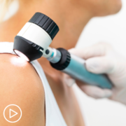
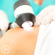
Along with a standard physical exam, many dermatologists use a technique called dermoscopy (also known as dermatoscopy, epiluminescence microscopy [ELM], or surface microscopy) to see spots on the skin more clearly. The doctor uses a dermatoscope, which is a special magnifying lens and light source held near the skin. Sometimes a thin layer of alcohol or oil is used with this instrument. The doctor may take a digital photo of the spot.

Skin biopsy

If the doctor thinks a spot might be a melanoma, the suspicious area will be removed and sent to a lab to be looked at under a microscope. This is called a skin biopsy.

There are many ways to do a skin biopsy. The doctor will choose one based on the size of the affected area, where it is on your body, and other factors. Any biopsy is likely to leave at least a small scar. Different methods can result in different types of scars, so ask your doctor about scarring before the biopsy. No matter which type of biopsy is done, it should remove as much of the suspected area as possible so that an accurate diagnosis can be made.

Skin biopsies are done using a local anesthetic (numbing medicine), which is injected into the area with a very small needle. You will likely feel a small prick and a little stinging as the medicine is injected, but you should not feel any pain during the biopsy.

Shave (tangential) biopsy

For this type of biopsy, the doctor shaves off the top layers of the skin with a small surgical blade. Bleeding from the biopsy site is stopped by applying an ointment, a chemical that stops bleeding, or a small electrical current to cauterize the wound.

A shave biopsy is useful in diagnosing many types of skin diseases and in sampling moles when the risk of melanoma is very low. This type of biopsy is not generally used if a melanoma is strongly suspected unless the biopsy blade will go deep enough to get below the suspicious area. Otherwise, if it is a melanoma, the biopsy sample may not be thick enough to measure how deeply the cancer has invaded the skin.

Punch biopsy

For a punch biopsy, the doctor uses a tool that looks like a tiny round cookie cutter to remove a deeper sample of skin. The doctor rotates the punch biopsy tool on the skin until it cuts through all the layers of the skin. The sample is removed and the edges of the biopsy site are often stitched together.

Excisional and incisional biopsies

To examine a tumor that might have grown into deeper layers of the skin, the doctor may use an excisional (or less often, an incisional) biopsy.

  • An excisional biopsy removes the entire tumor (along with a small margin of normal skin around it). This is usually the preferred method of biopsy for suspected melanomas if it can be done, although this isn’t always possible.
  • An incisional biopsy removes only a portion of the tumor.

For these types of biopsies, a surgical knife is used to cut through the full thickness of skin. A wedge or sliver of skin is removed for examination, and the edges of the cut are usually stitched together.

“Optical” biopsies

Some newer types of biopsies, such as reflectance confocal microscopy (RCM), can be done without needing to remove samples of skin. To learn more, see What’s New in Melanoma Skin Cancer Research?

Biopsies of melanoma that may have spread

Biopsies of areas other than the skin may be needed in some cases. For example, if melanoma has already been diagnosed on the skin, nearby lymph nodes may be biopsied to see if the cancer has spread to them.

Rarely, biopsies may be needed to figure out what type of cancer someone has. For example, some melanomas can spread so quickly that they reach the lymph nodes, lungs, brain, or other areas while the original skin melanoma is still very small. Sometimes these tumors are found with imaging tests (such as CT scans) or other exams even before the melanoma on the skin is discovered. In other cases, they may be found long after a skin melanoma has been removed, so it’s not clear if it’s the same cancer.

In still other cases, melanoma may be found somewhere in the body without ever finding a spot on the skin. This may be because some skin lesions go away on their own (without any treatment) after some of their cells have spread to other parts of the body. Melanoma can also start in internal organs, but this is very rare, and if melanoma has spread widely throughout the body, it may not be possible to tell exactly where it started.

When melanoma has spread to other organs, it can sometimes be confused with a cancer starting in that organ. For example, melanoma that has spread to the lung might be confused with a primary lung cancer (cancer that starts in the lung).

Special lab tests can be done on the biopsy samples that can tell whether it is a melanoma or some other kind of cancer. This is important because different types of cancer are treated differently.

Biopsies of suspicious areas inside the body often are more involved than those used to sample the skin.

Fine needle aspiration (FNA) biopsy

FNA biopsy is not used on suspicious moles. But it may be used, for example, to biopsy large lymph nodes near a melanoma to find out if the melanoma has spread to them.

For this type of biopsy, the doctor uses a syringe with a thin, hollow needle to remove very small pieces of a lymph node or tumor. The needle is smaller than the needle used for a blood test. A local anesthetic is sometimes used to numb the area first. This test rarely causes much discomfort and does not leave a scar.

If the lymph node is just under the skin, the doctor can often feel it well enough to guide the needle into it. For a suspicious lymph node deeper in the body or a tumor in an organ such as the lung or liver, an imaging test such as ultrasound or a CT scan is often used to help guide the needle into place.

FNA biopsies are not as invasive as some other types of biopsies, but they may not always collect enough of a sample to tell if a suspicious area is melanoma. In these cases, a more invasive type of biopsy may be needed.

Surgical (excisional) lymph node biopsy

This procedure can be used to remove an enlarged lymph node through a small incision (cut) in the skin. A local anesthetic (numbing medicine) is generally used if the lymph node is just under the skin, but the person may need to be sedated or even asleep (using general anesthesia) if the lymph node is deeper in the body.

This type of biopsy is often done if a lymph node’s size suggests the melanoma has spread there but an FNA biopsy of the node wasn’t done or didn’t find any melanoma cells.

Sentinel lymph node biopsy

If melanoma has been diagnosed and has any concerning features (such as being at least a certain thickness), a sentinel lymph node biopsy (SLNB) is often done to see if the cancer has spread to nearby lymph nodes, which in turn might affect treatment options. This test can be used to find the lymph nodes that are likely to be the first place the melanoma would go if it has spread. These lymph nodes are called sentinel nodes (they stand sentinel, or watch, over the tumor, so to speak).

To find the sentinel lymph node (or nodes), a doctor injects a small amount of a radioactive substance into the area of the melanoma. After giving the substance time to travel to the lymph node areas near the tumor, a special camera is used to see if it collects in one or more sentinel lymph nodes. Once the radioactive area has been marked, the patient is taken for surgery, and a blue dye is injected in the same place the radioactive substance was injected. A small incision is then made in the marked area, and the lymph nodes are then checked to find which one(s) became radioactive and turned blue. These sentinel nodes are removed and looked at under a microscope.

If there are no melanoma cells in the sentinel nodes, no more lymph node surgery is needed because it is very unlikely the melanoma would have spread beyond this point. If melanoma cells are found in the sentinel node, the remaining lymph nodes in this area are typically removed and looked at as well. This is known as a lymph node dissection (see Surgery for Melanoma Skin Cancer).

If a lymph node near a melanoma is abnormally large, a sentinel node biopsy probably won’t be needed. The enlarged node is simply biopsied.

Lab tests of biopsy samples

Samples from any biopsies will be sent to a lab, where a doctor called a pathologist will look at them under a microscope for melanoma cells. Often, skin samples are sent to a dermatopathologist, a doctor who has special training in looking at skin samples.

If the doctor can’t tell for sure if melanoma cells are in the sample just by looking at it, special lab tests will be done on the cells to try to confirm the diagnosis. These might include:

  • Immunohistochemistry (IHC)
  • Fluorescence in situ hybridization (FISH)
  • Comparative genomic hybridization (CGH)
  • Gene expression profiling (GEP)

If melanoma is found in the samples, the pathologist will look at certain important features such as the tumor thickness and mitotic rate (the portion of cells that are actively dividing). These features help determine the stage of the melanoma (see Melanoma Skin Cancer Stages), which in turn can affect treatment options and prognosis (outlook).

Testing for gene changes

For some people with melanoma, biopsy samples may be tested to see if the cells have mutations (changes) in certain genes, such as the BRAF gene. About half of melanomas have BRAF mutations. Some drugs used to treat advanced melanomas are only likely to work if the cells have BRAF mutations (see Targeted Therapy for Melanoma Skin Cancer), so this test is important in helping to determine treatment options. Tests for changes in other genes, such as C-KIT, might be done as well.

A newer lab test known as DecisionDx-Melanoma looks at certain gene expression patterns in melanoma cells to help show if early-stage melanomas are likely to spread. This might be used to help determine treatment options. To learn more, see What’s New in Melanoma Skin Cancer Research?

Imaging tests

Imaging tests use x-rays, magnetic fields, or radioactive substances to create pictures of the inside of the body. They are used mainly to look for the possible spread of melanoma to lymph nodes or other organs. These tests are not needed for most people with very early-stage melanoma, which is very unlikely to have spread.

Imaging tests can also be done to help determine how well treatment is working or to look for possible signs of cancer coming back (recurring) after treatment.

Chest x-ray

This test might be done to help determine if melanoma has spread to the lungs, although a CT scan of the chest (see below) is often done instead.

Ultrasound

Ultrasound uses sound waves to create images of the inside of your body on a computer screen. This test might be used to look at the lymph nodes near the tumor, especially if it’s not clear if they’re enlarged based on a physical exam. Ultrasound is typically fairly quick and easy to do, and it doesn’t expose you to radiation.

Ultrasound-guided needle biopsy: Ultrasound can also be used to help guide a biopsy needle into a suspicious lymph node.

Computed tomography (CT) scan

The CT scan uses x-rays to make detailed, cross-sectional images of your body. Unlike a regular x-ray, CT scans can show the detail in soft tissues (such as internal organs). This test can show if any lymph nodes are enlarged or if organs such as the lungs or liver have suspicious spots, which might be from the spread of melanoma.

CT-guided needle biopsy: CT scans can also be used to help guide a biopsy needle into a suspicious area within the body.

Magnetic resonance imaging (MRI) scan

MRI scans use radio waves and strong magnets instead of x-rays to create detailed images of parts of your body. MRI scans can be very helpful in looking at the brain and spinal cord.

Positron emission tomography (PET) scan

PET scan can help show if the cancer has spread to lymph nodes or other parts of the body. It is most useful in people with more advanced stages of melanoma.

For this test, you are injected with a slightly radioactive form of sugar, which collects mainly in cancer cells. A special camera is then used to create a picture of areas of radioactivity in the body.

PET/CT scan: Many centers have special machines that do both a PET and CT scan at the same time (PET/CT scan). This lets the doctor compare areas of higher radioactivity on the PET scan with the more detailed appearance of that area on the CT scan.

Blood tests

Blood tests aren’t used to diagnose melanoma, but some tests may be done before or during treatment, especially for more advanced melanomas.

Doctors often test blood for levels of a substance called lactate dehydrogenase (LDH) before treatment. If the melanoma has spread to distant parts of the body, a high LDH level is a sign that the cancer may be harder to treat. This can affect the stage of the cancer (see Melanoma Skin Cancer Stages).

Other tests of blood cell counts and blood chemistry levels may be done in a person who has advanced melanoma to see how well the bone marrow (where new blood cells are made), liver, and kidneys are working before and during treatment.

The American Cancer Society medical and editorial content team

Our team is made up of doctors and oncology certified nurses with deep knowledge of cancer care as well as journalists, editors, and translators with extensive experience in medical writing.

Last Medical Review: August 14, 2019 Last Revised: August 14, 2019


It’s NOT Just Skin Cancer…

Real patient experiences shared privately at www.TreatmentDiaries.com.  Read more, share if you like or join in the conversation.  Making sure you feel less alone navigating a diagnosis is important.  Connecting you to those who can relate and provide support is what we do.

I’m new to Treatment Diaries and since this is my diary, I want to share some thoughts that are now near and dear to my personal experience with this dreadful condition.  Let me start with what I’ve heard more times than I can count over the past decade and most often when sharing my Melanoma diagnosis with those who are uninformed.  It goes something like this – “What kind of cancer did you have?”  My response, “I was diagnosed with stage III Melanoma.”  The exchange – “Oh I think I’ve heard of that, it’s just skin cancer…right?”  In fact, I’ve had people tell me I was lucky to just have skin cancer.  Quite possibly the one thing you should consider never saying to someone with Melanoma.  Not only is it completely untrue it will do nothing to make the individual with the diagnosis feel any better about their situation.  The truth is, Melanoma is one of the deadliest forms of skin cancer.  It’s not just skin cancer.

So now that we are clear on it’s not just skin cancer, a few things I wish I would have known:

  • Research suggests that approximately 90% of melanoma cases can be linked to exposure to ultraviolet (UV) rays from natural or artificial sources, such as sunlight and indoor tanning beds.
  • However, since melanoma can occur in all melanocytes throughout the body, even those that are never exposed to the sun, UV light cannot be solely responsible for a diagnosis, especially mucosal and ocular melanoma cases.
  • Current research points to a combination of family history, genetics and environmental factors that are also to blame.
  • You can read this Melanoma Fact Sheet for more information!
  • Support for melanoma patients is incredibly important and connecting with those who relate brings much needed encouragement along with valuable insight.

Unlike other cancers, melanoma can often be seen on the skin, making it easier to detect in its early stages. Keeping track of the changes to your skin and seeing a dermatologist on an annual basis can be a lifesaving event. If left undetected, however, melanoma can spread to distant sites or distant organs. Once melanoma has spread to other parts of the body (known as stage IV), it is referred to as metastatic, and is very difficult to treat. In its later stages, melanoma most commonly spreads to the liver, lungs, bones and brain; at this point, the prognosis is very poor.  Again…it’s not just skin cancer.

Skin cancer comes in many forms and for numerous reasons.  Your job is to protect your skin.  Our skin is the biggest most vital organ we have to care for.  We can’t live without it nor can it be replaced.  It’s ours for as long as we live so we need to take the vitality of it seriously.  Stay out of the sun, look for changes and recurring issues to your skin even in areas that never see the sun, see a dermatologist on a regular basis and make sure to wear sunscreen all year around.  It’s not just skin cancer especially when it can kill you.  It’s a serious topic and I feel so very fortunate to be able to share my personal experience.  I hope to be a help to others newly diagnosed and an inspiration to those on the journey.  Let’s kick melanoma to the curb together…

Tag Archive for: skin cancer

Coming Soon

Please check back soon as we work to build more resources.